Physical Address
304 North Cardinal St.
Dorchester Center, MA 02124

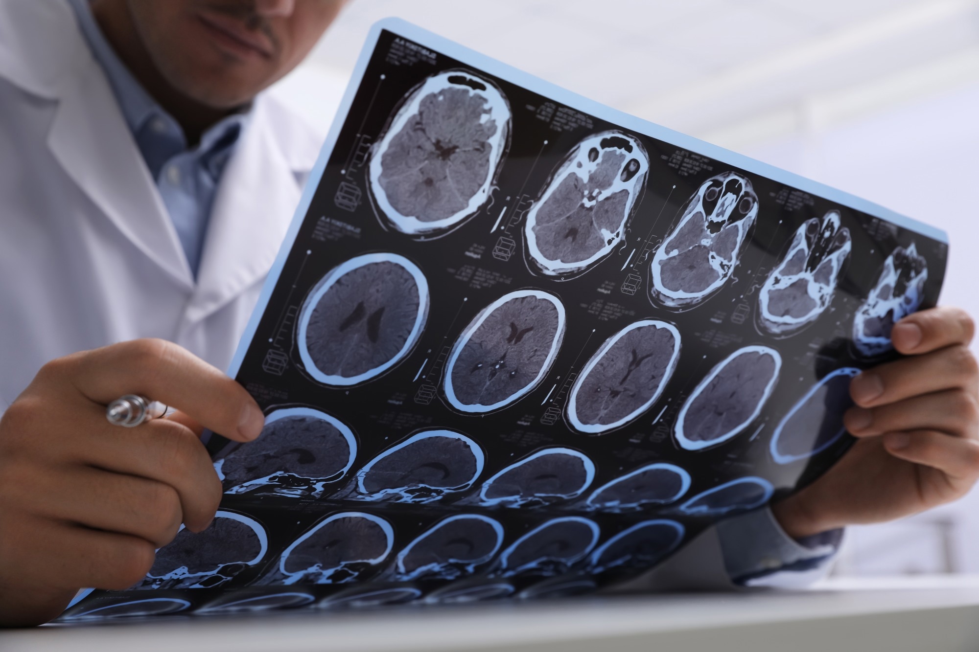
Neuroimaging is a pivotal tool for early dementia diagnosis, enabling visualization of brain changes. It aids in identifying cognitive disorders before pronounced symptoms develop.
Neuroimaging technologies, such as MRI and PET scans, have revolutionized the early detection and diagnosis of dementia, particularly Alzheimer’s disease. These advanced techniques allow doctors to see detailed images of the brain, detecting abnormalities like atrophy or amyloid plaques that may indicate the onset of dementia.
With early diagnosis, patients can access treatment and support sooner, potentially slowing the progression of the disease. Understanding the role of neuroimaging in dementia care is crucial for patients, caregivers, and healthcare professionals alike. It represents a significant step in managing the challenges of a disease that affects millions worldwide.
Dementia often starts as a whisper of forgetfulness before growing into a chorus of cognitive challenges. Spotting this condition in its early stages is crucial. Yet, early detection presents a labyrinth of obstacles. Today, we venture into the intricate world of dementia, unraveling the threads of its types, symptoms, and the uphill battle of early diagnosis.
Dementia is not a single disease but a term that encompasses a range of cognitive impairments. Here are some common types:
Each type of dementia comes with its own set of symptoms. Memory lapses, trouble finding words, and changes in mood are early indicators that shouldn’t be ignored.
Catching dementia in its infancy is pivotal. Early intervention can slow the progression and enhance quality of life. It allows:
Effective management hinges on recognizing the warning signs swiftly and accurately.
The traditional path to diagnosing dementia often relies on cognitive tests and medical history analysis. However, these methods can miss early subtle signs.
Contrastingly, neuroimaging represents a beacon of hope. Cutting-edge tools like MRI and PET scans illuminate the brain’s functioning, offering a glimpse into the early onset of dementia. This technological leap forward provides unparalleled precision in detection.
Early detection of dementia is critical for treatment and care planning. Neuroimaging technologies offer a window into the brain’s workings, helping to identify signs of dementia before symptoms fully emerge. This section delves into the main imaging techniques used to visualize the brain’s structure and function during the early stages of dementia.
Structural imaging provides detailed pictures of the brain’s anatomy. This reveals brain volume and detects changes that could suggest dementia.
Functional imaging assesses brain activity. It detects how well different brain regions are working.
Newer techniques offer deeper insights into brain health and dementia progression.
Every neuroimaging technique has its pros and cons. Each offers unique information about the brain.
| Technique | Main Use | Strengths | Limitations |
|---|---|---|---|
| MRI | Brain structure | Detail, no radiation | Cost, time |
| CT | Quick scanning | Accessibility | Less detailed |
| PET | Metabolism analysis | Detailed function | Radiation exposure |
| SPECT | Blood flow | Function insight | Less resolution |
| fMRI | Activity tracking | Real-time data | Complexity |
| DTI | White matter mapping | Damage detection | Less common |
Neuroimaging stands at the forefront of modern medical techniques aiming to unveil the early signs of dementia. Detecting cognitive diseases in their infancy stages, neuroimaging helps prepare for the onset of dementia. Approaches like MRI and PET scans reveal critical insights into brain health, facilitating early intervention strategies.
Neuroimaging techniques such as Magnetic Resonance Imaging (MRI) and Positron Emission Tomography (PET) scans provide a glimpse into the brain’s anatomy. They highlight changes like shrinkage in hippocampal regions, often signalling dementia. Functional MRI (fMRI) sheds light on how brain regions communicate, marking deviations from normal brain activity.
Accurate diagnosis is crucial as dementia symptoms resemble other medical issues. Neuroimaging helps differentiate dementia from conditions with similar symptoms, ensuring appropriate treatment. For instance, MRI scans differentiate vascular dementia from Alzheimer’s by identifying blood flow problems in the brain.
Neuroimaging has transformative potential for those with a family history of dementia. Brain scans in asymptomatic individuals reveal potential risk even before symptoms surface. This early window provides a chance for preventive measures to delay or hinder disease progression.

Credit: www.news-medical.net
Dementia often emerges silently, affecting memory, thought processes, and social abilities. Neuroimaging is a critical tool for early detection. Yet, this technology faces limitations and ethical dilemmas. Understanding these barriers enables better diagnostic practices and heightened ethical standards.
Current neuroimaging techniques are not flawless. Magnetic Resonance Imaging (MRI) and Positron Emission Tomography (PET) are common yet have their own drawbacks. MRI struggles to detect early subtle changes associated with dementia. PET scans, while more sensitive, are pricey and limited to certain locations.
The benefits of neuroimaging are clear, but not everyone can access these services. Factors such as location, availability, and insurance coverage must change. Investments in cost-effective solutions are crucial.
Early diagnosis of dementia raises sensitive concerns. Identifying dementia early means patients must confront potential stigma and discrimination. Healthcare institutions must establish protocols to safeguard privacy and provide support. They must also communicate risks and benefits clearly to those affected.
| Protocol Area | Response Strategy |
|---|---|
| Privacy | Maintain confidentiality, secure data |
| Support | Offer counseling, resources for planning |
Continuous research is pivotal. Scientists and policymakers must collaborate to enhance neuroimaging technologies. This collaboration should aim to make early dementia diagnosis more accurate and widely accessible. Ongoing policy work can then ensure that ethical practices keep pace with these advancements.
Neuroimaging techniques are revolutionizing dementia diagnosis. Detailed case studies and clinical evidence show how these methods can detect early signs of the condition. Identifying dementia early can be vital for effective intervention, improving patient outcomes.
Many patients now live better lives due to early dementia detection using neuroimaging. MRI and PET scans can reveal brain changes that suggest dementia. Early treatment plans can then be crafted. These success stories fuel the drive for wider neuroimaging use.
Despite successes, neuroimaging’s predictive accuracy is under scrutiny. Cases exist where scans predicted dementia that didn’t develop. Robust clinical trials are crucial to gauge the true accuracy of these methods.
| Type of Scan | Predictive Accuracy |
|---|---|
| MRI | Varied results across different studies |
| PET | Higher accuracy in certain dementia types |
Not all neuroimaging results lead to correct diagnosis. Misdiagnoses can result from over-relying on scans without considering clinical signs. Studies of these missteps help refine diagnosis procedures and caution against premature conclusions.
Different countries use neuroimaging in diverse ways due to resource availability and medical guidelines. Emerging markets may prioritize different techniques, influencing global dementia diagnosis strategies.
| Country | Preferred Neuroimaging Technique |
|---|---|
| USA | Advanced PET scans |
| India | Basic MRI scans |

Credit: www.neurology.org
The journey of understanding dementia has led to the doorstep of advanced diagnostic tools. Neuroimaging stands out as a beacon of hope for early detection. With this tool, the future in battling dementia unfolds with a new narrative of precision and timeliness.
Neuroimaging has transformed dementia diagnosis. It offers clear images of the brain’s structure and its workings. This breakthrough allows doctors to see the early signs of dementia. It shows the changes in the brain before symptoms get worse. Accurate early diagnosis can lead to better care for patients.
This progress means doctors may soon have more tools to spot dementia early.
Integration of neuroimaging in everyday care is on the horizon.
This shift will ensure all patients benefit from these advances.

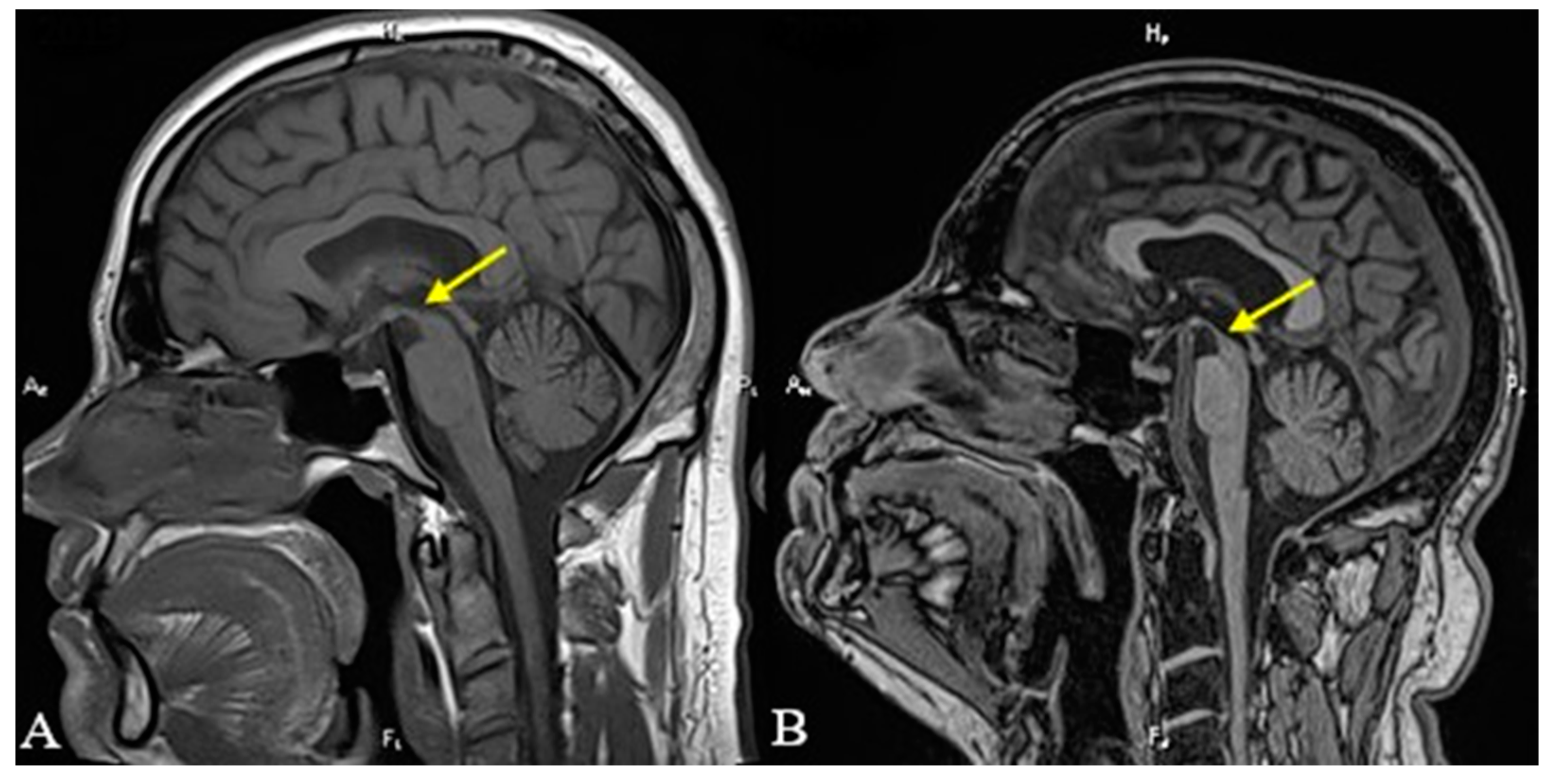
Credit: www.mdpi.com
Brain scans can reveal signs of early dementia by identifying brain structure changes and function impairments commonly associated with the condition.
Neuroimaging aids in diagnosing dementia by revealing brain structure and function changes, assisting doctors in identifying patterns consistent with various dementia types. It helps differentiate between dementia causes and gauges disease progression.
Upon early onset dementia diagnosis, consult a specialist for a treatment plan. Seek support from dementia-related groups, organize legal affairs, and consider future caregiving options. Stay healthy through diet and exercise, and keep mentally active.
Yes, medical professionals can use cognitive and neurological exams to assess for early signs of dementia. These tests evaluate memory, problem-solving, and other brain functions.
Neuroimaging stands at the forefront of revolutionizing early dementia detection. Its advanced techniques offer hope for timely interventions, potentially slowing disease progression. Embracing such technology could transform patient outcomes and care strategies. As research evolves, the promise of better diagnostics and improved quality of life for those affected by early-stage dementia becomes increasingly tangible.
Let’s advocate for continued innovation in this critical field of medicine.

